
Automatic image analysis is a step closer, thanks to new software capable of automatically tracing the path of filamentary structures in medical images, such as blood vessels in the retina.
© Joshua Carpenter/iStock/Thinkstock
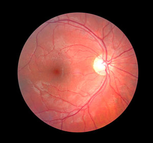
Automatic tracing of filamentary structures in medical images could improve drug screening and clinical diagnosis, and might also be useful for image analysis in other fields. Software to perform this complex task has been developed by A*STAR researchers in collaboration with co-workers in Singapore and China. The system has been used to examine images of neurons in brain tissue samples and blood vessels in the retina.
Clinical investigators often need to identify single neurons in images when assessing the effects of drugs being developed to treat neurological conditions such as Parkinson’s and Alzheimer’s diseases. Identifying the path of individual blood vessels in the back of the eye is used to diagnose damage caused by diabetes. Automating image analysis could make it faster and easier for clinicians to use, without requiring assistance from a technical expert. Some semi-automated systems already exist, but they frequently require human intervention to trace the complete paths of individual filaments in each image.
“A major bottleneck in automating the process has been the problem of filament crossovers,” says Li Cheng of the A*STAR Bioinformatics Institute. He explains it can be difficult for any automated system to trace a single filament past the points where they crossover or touch branches from neighboring neurons or blood vessels.
Cheng’s team, along with co-workers at other A*STAR Institutes, Nanyang Technological University in Singapore and Beijing Institute of Technology in China, tackled the problem with sophisticated image analysis techniques. In particular, a procedure called directed graph theory helped them trace the filamentary structures pixel by pixel. Their software has been tested on database images of neurons and retinas, which has confirmed that it could be developed into a high throughput automated system.
“Our tracing system significantly outperforms existing semi-automated procedures,” Cheng says, adding, “Having developed this powerful system we are now keen to deploy it to real-world challenges.” To pursue that aim the team want to highlight its possibilities to potential users worldwide. Patenting and commercialization will follow in due course if the initial promising indications are verified.

Li Cheng (front row, center) with colleagues.
© 2015 A*STAR Bioinformatics Institute
The software might also be applied to other medical and biological problems such as the movement of materials within cells. “We hope it will eventually lead to the faster identification of better drugs for saving patents’ lives,” says Cheng. He points out that it may also be able to trace features in the built and natural environment, such as road networks.
The A*STAR-affiliated researchers contributing to this research are from the Bioinformatics Institute, the Institute of Molecular and Cell Biology and the Institute of Medical Biology. For more information about the team’s research, please visit the Machine Learning For Bioimage Analysis webpage.


