
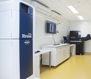
The A*STAR Singapore Bioimaging Consortium has partnered with Bruker Corporation to establish a Preclinical Imaging Centre offering training, demonstration and research for two cutting-edge imaging technologies: In-Vivo Xtreme and Skyscan 1176.
© 2015 Bruker Corporation
The A*STAR Singapore Bioimaging Consortium (SBIC) has partnered with scientific instrument manufacturer Bruker Corporation to offer researchers in Singapore access to some of the most advanced imaging technologies for preclinical research. “The SBIC–Bruker Preclinical Imaging Centre is one of a series of investments that Bruker is making in our core business,” says Nikita Agarwal, applications specialist at Bruker.
Launched in October 2014, the center provides training, demonstration and research facilities, which support the development of preclinical imaging applications customized to researchers’ needs in emerging fields. Pertinent examples include the translation of SBIC’s wide repertoire of fluorescence probes into clinically relevant biomarkers and the identification of anatomical and molecular changes in animal models of disease.
“This is a mutually beneficial partnership,” says Young-Tae Chang who heads the SBIC Laboratory of Bioimaging Probe Development. “We provide a cutting-edge, user perspective to further enhance Bruker’s machines, while accelerating our research toward a clinical direction.”
Chang’s team has produced the most extensive fluorescence probe toolbox in the world, with probes that can specifically tag and track a wide range of biomarkers, including stem cells, macrophages and, most recently, neurons. Fluorescent probes are ideal for studying biological processes in small animals at shallow depths of up to several millimeters. However, as light sources cannot excite the probes at greater depths in the human body, Chang plans to use radioactive versions that emit gamma rays, which can be detected using positron emission tomography and single-photon emission computed tomography cameras. Using the broad-spectrum detection capabilities of In-Vivo Xtreme, his team will be able to accelerate this characterization process.
“Bruker offers nine preclinical imaging modalities,” says Agarwal. “Within each piece of equipment, we try to include as many features as possible to give researchers more bang for their buck.”
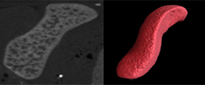
Skyscan 1176 can be used to reconstruct high-resolution, three-dimensional models of the mouse spleen (right) that enable closer investigation of anatomical structures than conventionally obtained grayscale images (left).
© 2015 Bruker Corporation
While probe developers may lean toward Xtreme (a multimodality measurement system for fluorescence, bioluminescence, X-ray and radioisotopes), researchers measuring effects at high resolution would be partial to the micro-computed tomography system Skyscan 1176, explains Agarwal. Skyscan uses X-rays to generate high-resolution, three-dimensional images of anatomical structures inside the body with resolutions of up to 9 micrometers. “It is the equivalent of a noninvasive in vivo microscope,” she says. The system can be used to study tumor formation or bone regeneration applicable to new fields such as stem cell therapy. Employed in combination with contrast agents, which visually enhance finer structures, Skyscan can be used to investigate soft tissues such as fat, blood vessels and individual organs.
The imaging technologies developed by the center complement those already available at SBIC such as magnetic resonance imaging. Moreover, it also adds to SBIC-led initiatives aimed at closer engagement with industry — which include the SBIC–Nikon Imaging Centre and more than 20 projects agreed under the Translational Imaging Industrial Laboratory providing preclinical imaging resources and solutions to pharmaceutical companies. “The SBIC is seen as a national resource in Singapore that serves not only A*STAR but also the wider community through universities and hospitals,” says Kishore Bhakoo who directs the industrial laboratory.
“SBIC is well regarded in Asia for imaging and is on its way to being recognized globally in this field,” adds Agarwal. “We felt that it made an excellent partner for our Preclinical Imaging Centre in Asia.”
About A*STAR’s Singapore Bioimaging Consortium
The Singapore Bioimaging Consortium (SBIC) is a research consortium of the Agency for Science, Technology and Research (A*STAR) in Singapore. SBIC aims to build a coordinated national programme for imaging research, bringing together substantial strengths in the physical sciences and engineering and those in the biomedical sciences. It seeks to identify and consolidate the various bioimaging capabilities across local research institutes, universities and hospitals, in order to speed the development of biomedical research discoveries.
SBIC has built up an extensive network of partners, through collaborations and joint appointments with research institutes, university and clinical departments, and the industry. Its partners include the Singapore Institute for Clinical Sciences, the NUS Chemistry Department, the National Cancer Centre, Duke-NUS, Agilent, Bruker, Bayer, GlaxoSmithKline, Takeda, and Merck.


