
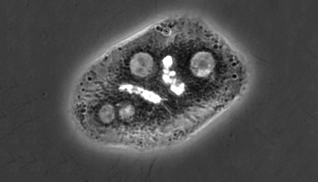
A phase contrast image which shows three hepatocytes forming two epithelial tubes (bright tubular structures).
© 2016 A*STAR Institute of Bioengineering and Nanotechnology
Researchers from Singapore and France have demonstrated, for the first time, that the mechanical forces generated by surrounding ‘scaffolding’ help to shape some types of epithelial tubes.
Epithelial tubes form much of the plumbing of our bodies. These tubes are found in all the major organs and allow the flow of fluids that sustain life. For example, the liver contains an extensive network of epithelial tubes that distribute bile acids — compounds that enable the digestion and absorption of fats in the small intestine. Defects in the formation of these tubes can lead to diseases such as cholestasis, in which the flow of bile is impeded or stopped.
One aspect that has mystified biologists is how such long, elongated tubes form when most developmental processes operate equally in all directions to produce symmetric structures, rather than the asymmetric tubular structures of epithelial tubes.
Now, a team led by Virgile Viasnoff at the National University of Singapore and CNRS (France) and Hanry Yu at the A*STAR Institute of Bioengineering and Nanotechnology has shown that forces produced by supporting cells — generated by the asymmetric interaction with the extracellular matrix — give rise to the long cellular architectures of some epithelial tubes.
“This is an exciting discovery since it means we can obtain epithelial tubes of different shapes and sizes by manipulating the microenvironment of these cells,” says Yu.
Adopting a minimalist approach the team experimented by culturing just two liver cells in tissue culture. When these cells were grown between walls coated with extracellular matrix, the three-dimensional arrangement of the extracellular matrix affected the shape of the lumens (the ‘holes’ in the cells that form the epithelial tubes). In particular, the lumens tended to move away from the extracellular matrix and toward the cell–cell interface, giving rise to the tubular structure.
The researchers modeled the situation mathematically and found that this elongation was driven by forces generated by the asymmetric interaction between cells and the extracellular matrix.
“Our study has revealed for the first time the exact nature of this mechanism and how it works,” says Yu. “It will contribute to a deeper understanding of the principles underlining epithelial tube formation as well as offer opportunities to develop better therapies for related diseases.
The team is keen to explore the medical implications of their discovery. “My group is focusing on translating our findings to different applications,” says Yu. “Specifically, we are studying the mechanism of tube dynamics and developing assays to screen for cholestasis.”
The A*STAR-affiliated researchers contributing to this research are from the Institute of Bioengineering and Nanotechnology and the Institute of Molecular and Cell Biology.


