
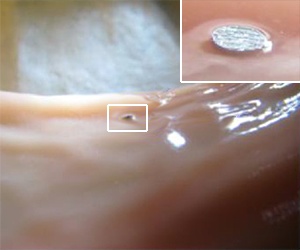
The microtag embedded in pig intestine
© 2011 IOP
Capsule endoscopes are powerful tools for the diagnosis of gastrointestinal (GI) disorders. These tiny wireless bioimaging devices, which are swallowed with a sip of water, scan the digestive system and send images remotely to an external workstation as they travel through the body. Vaidyanathan Kripesh and co-workers at the A*STAR Institute of Microelectronics have now developed a heat-activated tagging module that detects and labels abnormal tissue along the GI tract for these endoscopes.
There are many methods currently available for estimating the location of abnormal tissue or lesions. Some methods determine the approximate lesion location from the speed and transit times of the capsule as it traverses the stomach and small intestine, while other methods rely on the estimated path length of the capsule along the GI tract or the correlation between signal intensity levels detected by sensors positioned around the body and offline captured images. However, these approaches are unable to determine the exact position of lesions, delaying treatment and causing patient’s discomfort.
To speed up diagnosis and treatment, the researchers engineered a tagging module that can be integrated into conventional capsules. “The main limitations to incorporating an additional feature into capsules were size restriction and power consumption,” explains Kripesh. These constraints prompted them to design a compact low-power unit composed of a needle-sharp microscopic tag to label tissue, and a heat-triggered actuator to hold and release the tag.
The researchers manufactured the tag using titanium, a biocompatible material commonly used for X-ray imaging. The actuator consists of a piston and a helix-shaped spring maintained in a compressed state using a nylon thread latch that is in direct contact with a small silicon heater. Ignition of this heater breaks the latch, causing the spring to expand and propel the tag out of the module.
This approach allows physicians to trigger the module wirelessly when identifying the area of interest during imaging, with the entire process taking less than 30 seconds. “The doctor can use X-ray imaging for a faster localization,” says Kripesh.
The researchers assessed the performance of the tagging module through ex vivo experiments on pig intestine specimens. The heat-induced breakage of the latch immediately released the tag, which penetrated the tissue regardless of orientation. X-ray analysis clearly showed the implanted tags.
The researchers are currently miniaturizing the module so that the capsule can house four or more modules for multi-site labeling. They are also producing tags that the body can absorb for enhanced biocompatibility.
The A*STAR-affiliated researchers contributing to this research are from the Institute of Microelectronics.


