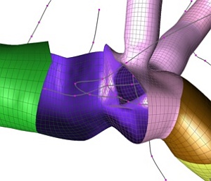
Illustration of a hole that was made according to a branch of the liver vascular system. The different parts of the combined mesh are displayed in different colors.
© Feiniu Yuan
Feiniu Yuan and co-workers at the A*STAR Singapore Bioimaging Consortium have developed a technique that automatically generates three-dimensional (3D) mesh models of the liver’s blood vessel system from medical images. The computer technology is beneficial not only for clinical diagnosis and surgery, but also for medical teaching and training.
Building an accurate computer model of liver vasculature has proved to be challenging in the past because of the small size of many of the vessels, the difficulty of gaining sharp images, and the fact that the system is highly branched. Yuan and co-workers overcame the problem by taking a ‘vessel tree modeling (VTM)’ approach.
Three-dimensional medical images are generated by tomography — a method that involves combining stacks of 2D images or sections. In these sections, tubular blood vessels appear circular, except at branch points. In their VTM approach, the researchers program a computer to track matching circular segments through sequences of sections from a designated root point, and also to note the branch points. The computer then generates centerlines of each vessel, by joining the centerpoints of sequential circular sections, and organizes these into a tree based on the branch point information.
The modeling technique then breaks the tree into its separate branches and forms a coarse mesh cylinder for each branch. It discards branches too small for the imaging technique to resolve sharply. At the branch points, the model fits the cylinders together, by generating a hole in the longer branch and fitting in and merging the base of the shorter branch. For multiple branches the procedure is repeated several times. The completed vessel tree structure is then smoothed using a fine mesh. The technique produces a model with relatively minimal error.
The new technique offers several unique advantages. Firstly, it generates a smooth mesh model that encompasses both large and small structures of the liver’s blood vessel system. Secondly, it is extremely efficient, enabling users to edit the mesh model in real-time.
In addition, given its capability in generating 3D mesh models of the liver’s blood vessel system, there is no reason why the technique cannot simulate the brain’s blood vessel system. The main limitations here, however, are that the technique can only model systems with a tree-like structure.
“Vascular modeling has many applications,” says Yuan. “Previous studies have shown that special shapes, such as regions with high curvature and bifurcations, are prone to the initiation and development of atherosclerosis. Our technique could provide scientists a deeper insight into the causes behind this.”
The A*STAR-affiliated researchers contributing to this research are from the Singapore Bioimaging Consortium.


