
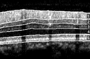
Fig. 1: Automatic segmentation of a spectral-domain OCT retinal image into five layers. The glaucoma can be diagnosed based on the thickness variation of the nerve fiber layer. 1, retinal nerve fiber layer; 2, ganglion cell layer; 3, inner plexiform/nuclear layer; 4, outer plexiform/nuclear layer; 5, photoreceptor layer.
© 2010 C.Y. Cheung
Optical coherence tomography (OCT) is an imaging technique that is being increasingly used to diagnose progressive eye conditions such as glaucoma. Shijian Lu and computer scientists at the A*STAR Institute for Infocomm Research have now teamed up with ophthalmologists to develop an automated method for analyzing OCT images.
OCT uses a light source with particular properties to generate cross-sectional images of translucent or opaque materials such as biological tissue. It does this by capturing light reflected by internal structures or discontinuities between tissue layers.
Glaucoma involves the loss of nerve cells from within the retinal nerve fiber layer (RNFL). This feature of the disease can be detected by measuring the RNFL thickness as well as that of other retinal layers from OCT images.
“Accurate and reliable automated analysis of OCT images should greatly increase the efficiency of diagnostic screening,” says Lu. Their method implements advanced spectral-domain OCT, which offers faster scanning, improved signal sensitivity and increased image definition. However, the automated segmentation of different anatomical layers within the retina from OCT images remains a challenging task.
Lu and his co-workers developed a system that automatically segments images generated by spectral-domain OCT into five retinal layers, including the RNFL (Fig. 1). “Our system is specifically designed for computer-aided diagnosis of glaucoma and other eye diseases, and has features not found in previously proposed methods for analyzing OCT images,” says Lu.
The new system first detects retinal blood vessels and then uses these to cut the image into multiple vessel and non-vessel sections. Based on the known retinal anatomy, it then detects boundaries within the non-vessel sections and classifies them into different retinal layers. Lastly, the retinal layer boundaries of the vessel sections are determined by interpolation, resulting in a complete picture of retinal boundary positions. Having determined the boundary positions, the thickness of the RNFL can be measured and quantified for glaucoma diagnosis and the monitoring of disease progression.
To test the performance of their proposed OCT segmentation technique, the researchers used it to analyze spectral-domain OCT images from four healthy subjects at the University Eye Center of the Chinese University of Hong Kong. They showed that that the technique accurately segments an OCT image into five layers, allowing the efficient assessment of RNFL thickness.
“We now intend to test the technique using larger-scale datasets and to introduce further improvements that are needed to allow reliable segmentation of layers much thinner than the RNFL,” says Lu.
The A*STAR-affiliated researchers contributing to this research are from the Institute for Infocomm Research.


