
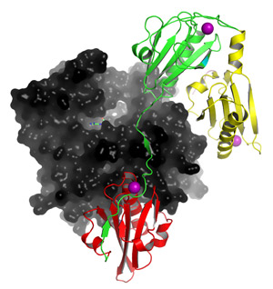
Fig. 1: Structure of a fragment of human gelsolin—containing domains G1 (red), G2 (green) and G3 (yellow)—bound to calcium (purple) and actin (black). Calcium binding in the G2 domain plays an essential role in initiating the structural rearrangements involved in protein activation.
Reproduced from Ref. 1 © 2009 National Academy of Sciences
Filaments of actin protein run throughout the interior of the cell, and contribute to diverse functions ranging from cell division and cell motility to the trafficking of essential molecules. These structures are highly dynamic, and their proper function depends on tightly regulated mechanisms for extension and disassembly, such as the filament-cleaving protein gelsolin.
Gelsolin features eight calcium ion-binding sites that contribute to protein activation, although many mechanistic details remain to be uncovered. “Gelsolin undergoes huge conformational changes in the process of becoming activated and then severing actin filaments,” says Robert Robinson of the A*STAR Institute of Molecular and Cell Biology, Singapore. “All of this is driven by simply binding a few calcium ions, and we want to know how this happens.”
Robinson’s team recently obtained high-resolution structural data for human gelsolin in its inactive calcium-free state as well as a large fragment of activated gelsolin (Fig. 1) bound to calcium and actin1. These two structures proved to be highly similar to structures previously obtained by his group for the equine protein2, but with a few notable differences—most importantly, the activated human protein was bound to an additional calcium ion at a site in its ‘G2’ domain.
This site, which has a functionally equivalent counterpart in a second domain (‘G6’), turns out to play a pivotal role in gelsolin activation. When these G2 and G6 calcium-binding sites are unoccupied, they interact and thereby contribute to stabilization of the highly compact inactive protein; calcium ion binding at G2 and G6 results in disruption of this association, followed by massive structural rearrangements. Mutational analysis of the other calcium-binding sites indicated that several of these may serve supporting roles in gelsolin activation.
Intriguingly, the mutation associated with the neuropathic disease familial amyloidosis Finnish type (FAF) is located in G2 at a position that disrupts calcium binding. Robinson and his co-workers predict that this change leaves the protein in an abnormal neither-activated-nor-inactivated conformation that is vulnerable to cleavage by the enzyme furin—thereby producing the insoluble clumps of truncated gelsolin that are a hallmark of FAF.
With this improved understanding of the role of calcium binding, Robinson’s team is now delving deeper into gelsolin function. “Our main goal is to determine the structure of gelsolin bound to more than one actin protomer to help us better understand the process of severing,” he says. In parallel, they also intend to test their model of FAF pathology and explore the potential for developing therapeutics that block furin cleavage.
The A*STAR-affiliated authors in this highlight are from the Institute of Molecular and Cell Biology.


