
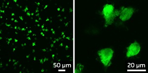
Fluorescent protein-tagged microglia in the brain of embryonic mice
Microglia are macrophages that reside in the central nervous system (CNS), constantly scavenging the brain for damaged neurons, plaque and foreign antigens. The CNS needs the right amount of microglial activity to maintain its wellbeing—too much or too little microglial activity could contribute to the development of neurodegenerative disorders such as Alzheimer’s and Parkinson’s disease.
Because microglia are so important to CNS health, scientists believe it might be possible to treat neurological disorders through the manipulation of microglia activity. In order to achieve this, however, it is important to understand microglial origins and regulation. A recent study by Florent Ginhoux and co-workers at the A*STAR Singapore Immunology Network has brought us closer to this goal.
Previous studies concerning the origins of microglia had drawn inconsistent conclusions. Ginhoux and his co-workers used innovative approaches to pinpoint where, when and how microglia arise in the CNS. Recent studies had found that microglial progenitors—the precursors of microglia—were already present in the developing brain at birth, but the researchers wanted to find out if these cells were definitive progenitors that will persist in the adult brain and how they came about during the development of the embryo.
To answer this, they used mice that express fluorescent proteins in immune cells and studied embryos of different ages to see when fluorescent cells could first be seen in the brain (see image). At embryonic day 9.5 of the 20 days of mouse development, they detected fluorescent cells not only in the developing brain but also in the yolk sac, which is known to give rise to the primitive macrophages present in early development. To test whether these yolk-sac primitive macrophages were the precursors of microglia, they used a type of mouse that can be made to irreversibly express fluorescence only within these yolk sac macrophages and their progeny. In this model, fluorescence was detected in adult microglial cells, showing unequivocally that microglia come from these yolk-sac progenitors.
These results are especially interesting because they show that microglia, as well as having specialized functions in the CNS, also have a unique origin. They are seeded into the brains of embryos by precursors from the embryonic yolk sac before the precursors of most types of macrophages have even developed.
“Our work reveals the unique origin of microglial cells in the immune system and that there are differentiation processes that only occur during early embryonic life. These results may lead to the development of new strategies for the treatment of various brain disorders,” says Ginhoux.
The A*STAR-affiliated researchers contributing to this research are from the Singapore Immunology Network.


