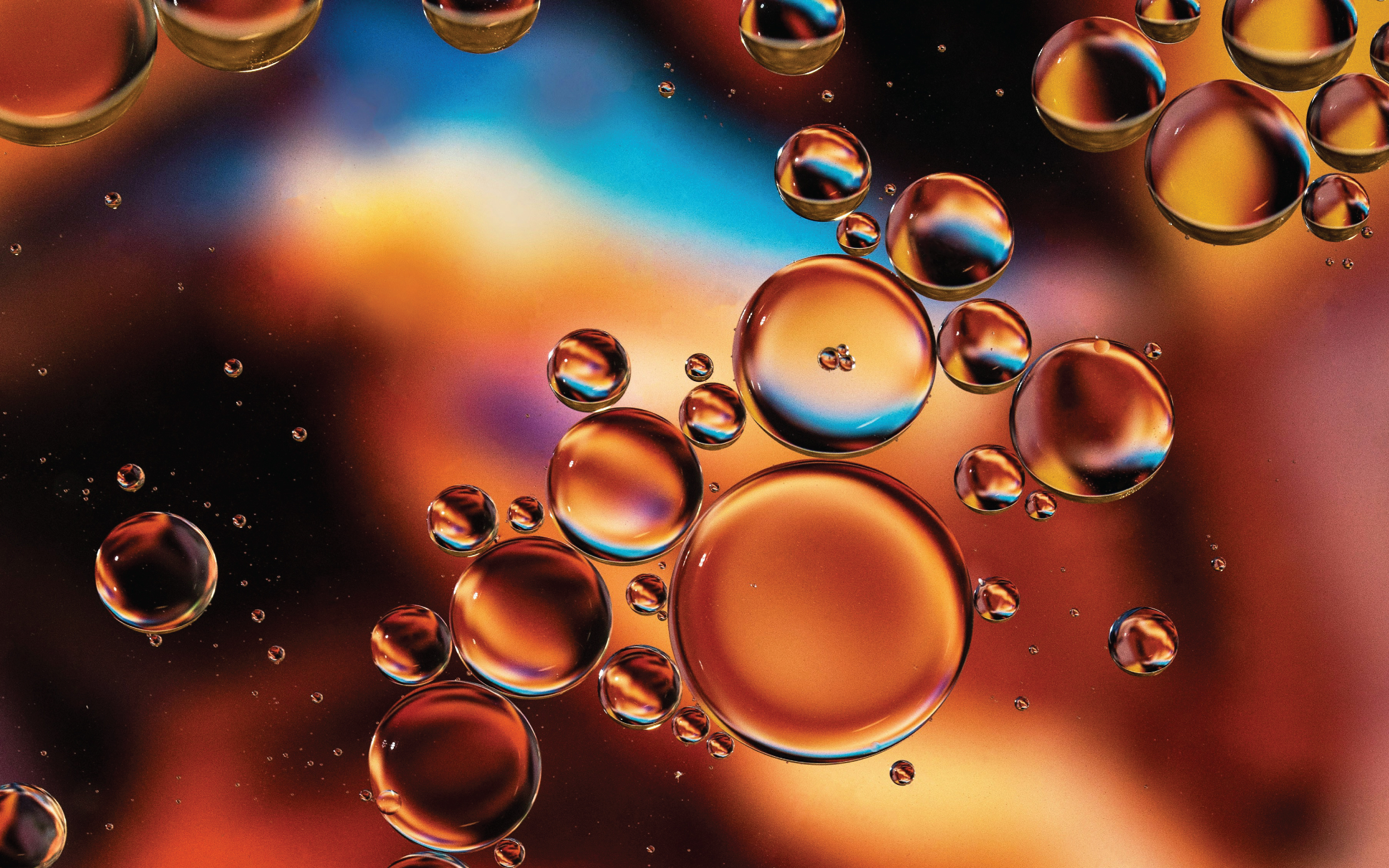
Viewed at the nanoscale, COVID-19 mRNA vaccines would appear as bubbles of fatty molecules, each encasing a nest of fine mRNA strands. These bubbles, also known as lipid nanoparticles, protect the delicate genetic instructions encoded by those strands, which would otherwise break down in the body within minutes.
When researchers design new treatments, drug delivery systems like lipid nanoparticles are crucial components to keep in mind, as they shield the actual therapeutic molecules long enough for them to reach the cells they’re meant for in one piece. However, Sangyong Jung, Group Leader at the A*STAR Institute of Molecular and Cell Biology (A*STAR IMCB), notes that designing such systems for mRNA therapies can be trickier than most, given how fragile mRNA can be.
“Lipid nanoparticles need precise manufacturing to keep mRNA stable and ensure it reaches the right cells,” said Jung. It’s a challenge compounded by the varied regulatory and production demands depending on the application; mRNA vaccines and mRNA gene therapies, for example, have very different optimal dosages and cellular targets.
Jung and colleagues partnered with Nam-Joon Cho’s group at Nanyang Technological University, Singapore, to investigate whether certain structural features seen in lipid nanoparticles could influence their stability and effectiveness when used as mRNA delivery systems.
“The surface properties and makeup of these lipid structures influence how quickly the mRNA-lipid complex breaks down,” Jung noted. “They also affect how the body’s immune system responds, which impacts how long the complex lasts and its chances of reaching its target cells or organs.”
Their research focused on spherical lipid nanoparticles called liposomes, as created through one of two methods: extrusion or the LUCA cycle, a novel freeze-thaw technique. By tweaking liposome shapes and adding short-chain lipids to them, the team found they could fine-tune their properties. Using cryo-electron microscopy (Cryo-EM) and other advanced imaging techniques, they observed how these modified liposomes interacted with mRNA.
The researchers found that the LUCA cycle produced ‘onion-like’ multi-layered structures, while the standard extrusion process generated simpler, single-layered liposomes. According to Jung, Cryo-EM showed that the multi-layered structures were particularly effective at securing mRNA, reducing the risk of it being released too early.
“The ‘onion-like’ structure is a potential indicator of quality during biophysical tests of the mRNA-lipid complex,” Jung added.
Through further studies in mice, the team confirmed that compared to simpler liposome structures, the multi-layered nanoparticles not only improved organ-specific targeting, but also the level of target gene expression. They also discovered that the liposomes' initial shape and structure was critical to achieving the right charge balance for effective mRNA delivery.
“Our findings provide valuable insights into the intricate relationship between lipid structures and the features of the resulting mRNA-lipid complexes, which can aid the development of mRNA delivery systems,” said Jung, “We’re currently exploring new lipid complexes that stabilise large mRNA in particular, as well as those that can home in on specific tissues.”
The A*STAR-affiliated researchers contributing to this research are from the A*STAR Institute of Molecular and Cell Biology (A*STAR IMCB).


